Home » Without Label » Back Muscle Diagram / Labeled Anatomy Chart Of Male Triceps And Back Muscles On White Background Stock Photo Download Image Now Istock / The part of the nerve that emerges out of the spine is called the nerve root.
Back Muscle Diagram / Labeled Anatomy Chart Of Male Triceps And Back Muscles On White Background Stock Photo Download Image Now Istock / The part of the nerve that emerges out of the spine is called the nerve root.
Back Muscle Diagram / Labeled Anatomy Chart Of Male Triceps And Back Muscles On White Background Stock Photo Download Image Now Istock / The part of the nerve that emerges out of the spine is called the nerve root.. They extend and rotate the head and neck. The back muscles enable you to stand up straight; In this image, you will find 1st cervical vertebrae, atlus, cervical plexus, 7th cervical vertebrae, 1st thoracic vertebrae, brachial plexus, spinal dura mater, filaments of spinal nerve roots, 12th thoracic vertebra, 1st lumber vertebra, iliohypogastric nerve, ilioinguinal nerve, lumbar. For more information visit www.threetreasuresstudio.com Muscles of the lower back and buttocks diagram, human muscles, muscles of the lower back and buttocks diagram.
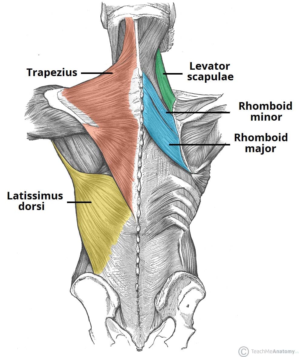
The back consists of the spine, spinal cord, muscles, ligaments, and nerves. Creatine is now proving to be one of the most potent muscle growth accelerators giving excellent muscle mass increase and phenomenal strength increases order yours today. A video describing the major muscles of the back. The trapezius is a broad, flat and triangular muscle. A strained muscle in your lower back can be quite painful.
Muscles Of The Back Teachmeanatomy from teachmeanatomy.info However, the spinal erectors travel the length of the entire spine. We did not find results for: Creatine research more than a sports supplement read more…. Maybe you would like to learn more about one of these? They extend and rotate the head and neck. A clip from 3d back muscles: Both the deltoid and the trapezius are firmly attached to the spine of the scapula. Diagrams of back muscles | 101 diagrams from www.101diagrams.com back muscle diagrams printable diagram.
For more anatomy content please follow us and visit our website:
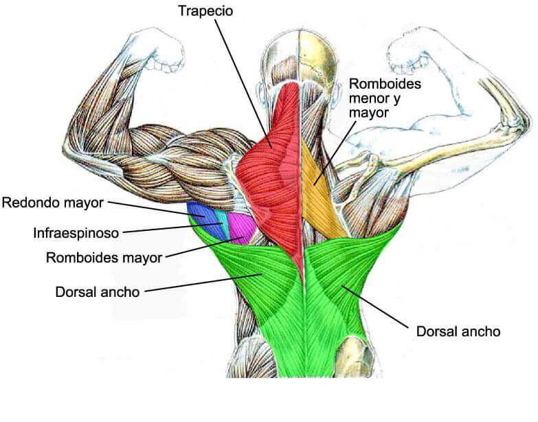
The fibres attach to the clavicle, acromion and the scapula spine. Major muscles back muscles shoulder muscles body anatomy human anatomy muscle chart anatomy human muscle anatomy supraspinatus muscle back workout routine. Muscles of the back diagram. See how exercise helps the back. What is the origin and insertion of the rhomboid minor and major muscle? Check spelling or type a new query. Muscles of the lower back diagram / male lower back muscles on black photograph by hank grebe. Saved by great big canvas. Anatomy of the spine and back spine muscles diagram. Superficial, intermediate, deep and deepest layers.these muscles lie on each side of the vertebral column, deep to the thoracolumbar fascia they span the entire length of the vertebral column, extending from the cranium to the pelvis For example, some muscles located in the chest also help move the shoulders. When back development is the goal, stick to one of these variations. Back muscles, back muscle diagram.
Superficial, intermediate, deep and deepest layers.these muscles lie on each side of the vertebral column, deep to the thoracolumbar fascia they span the entire length of the vertebral column, extending from the cranium to the pelvis However, the spinal erectors travel the length of the entire spine. Five pairs of lumbar spinal nerves labeled l1 to l5 branch off your spinal cord and exit through small holes between the vertebrae. The back consists of the spine, spinal cord, muscles, ligaments, and nerves. The part of the nerve that emerges out of the spine is called the nerve root.
Anatomy Of The Back Muscles Fit People from fitpeople.com We hope this picture anatomy of back muscles diagram can help you study and research. Maybe you would like to learn more about one of these? These structures work together to support the body, enable a range of movements, and send messages from the. However, the spinal erectors travel the length of the entire spine. For more anatomy content please follow us and visit our website: Major muscles back muscles shoulder muscles body anatomy human anatomy muscle chart anatomy human muscle anatomy supraspinatus muscle back workout routine. To learn more about the anatomy of the spine, watch this video. A strain can be an injury to a tendon attachment from muscle to bone.
Check spelling or type a new query.
Creatine research more than a sports supplement read more…. The pelvis at the bottom of the back and the shoulders at the top of the back give the back. Creatine is now proving to be one of the most potent muscle growth accelerators giving excellent muscle mass increase and phenomenal strength increases order yours today. We hope this picture anatomy of back muscles diagram can help you study and research. Some of the links in the post above are affiliate links.. In this image, you will find 1st cervical vertebrae, atlus, cervical plexus, 7th cervical vertebrae, 1st thoracic vertebrae, brachial plexus, spinal dura mater, filaments of spinal nerve roots, 12th thoracic vertebra, 1st lumber vertebra, iliohypogastric nerve, ilioinguinal nerve, lumbar. We think this is the most useful anatomy picture that you need. However, the spinal erectors travel the length of the entire spine. It is opposite from the chest, and the vertebral column runs down the back. They extend and rotate the head and neck. Another common cause of lower back and hip pain is disc injury. For example, some muscles located in the chest also help move the shoulders. See back muscles and low back pain.
The fibres attach to the clavicle, acromion and the scapula spine. While muscles like the gluteals (in the thighs) are used any time we walk or climb a step, deep back muscles and abdominal muscles are usually not actively engaged during everyday activity. For more information visit www.threetreasuresstudio.com The muscles on each side form a trapezoid shape. Deep back muscles diagram the superficial layer contains the splenius cervicis and splenius capitis muscles.
Low Back Muscles Anatomy Anatomy Drawing Diagram from i.pinimg.com A strain can be an injury to a tendon attachment from muscle to bone. We did not find results for: To learn more about the anatomy of the spine, watch this video. It is the most superficial of all the back muscles. We think this is the most useful anatomy picture that you need. Nerves in your lower back. Likewise, there are muscles in other parts of the body that help support and move the spine. When back development is the goal, stick to one of these variations.
A strained muscle in your lower back can be quite painful.
To learn more about the anatomy of the spine, watch this video. For more information visit www.threetreasuresstudio.com Your back hurting more when you move, less when you stay still; The human back extends from the buttocks to the posterior portion of the neck and shoulders. Muscles of the back diagram. This is a diagram of the larger and more surface muscles of the low back. The back consists of the spine, spinal cord, muscles, ligaments, and nerves. These are typical symptoms you might experience: Five pairs of lumbar spinal nerves labeled l1 to l5 branch off your spinal cord and exit through small holes between the vertebrae. Lower back muscle diagram : The back muscles enable you to stand up straight; When back development is the goal, stick to one of these variations. It is the most superficial of all the back muscles.